Tuesday, 19 March 2024 00:00
Congenital Foot Problems
A congenital foot problem is a problem affecting the feet, toes, and/or ankle that a child is born with. Several issues with a child’s feet can occur congenitally. Such problems include clubfoot, vertical talus, tarsal coalition, polydactyly, macrodactyly, and cleft foot. Some of these problems have a genetic basis, with someone in their family history having a gene causing the condition, and some are simply an anomaly.
The following are specifics about a few of these conditions:
- Clubfoot, also called congenital talipes equinovarus or talipes equinovarus, is When the tendons of the foot shorten, the bones are of an unusual shape, and the Achilles tendon is tight, causing an inward and downward pointing of the foot. The soles of the feet might also face each other. In most cases of clubfoot, both feet are affected. If not treated, the affected child will walk on the sides of their feet or ankles.
- Polydactyly is a condition where the child has more than five fingers or toes on either or both feet. Presentation usually consists of a nubbin or small lump of tissue without a bone, a toe that is partially formed but has no joints, or an extra toe.
- Vertical talus is where the talus bone forms in the wrong position, other bones in the foot do not line up properly, the front of the foot points up, and the bottom of the foot is stiff, has no arch, and usually curves out. This can occur in one or both feet and if left untreated, can lead to serious disability or discomfort as the child grows.
- Tarsal coalition is when there is an abnormal connection of two or more bones in the foot leading to severe, rigid flatfoot. The tarsal bones, located toward the back of the foot and in the heel, are the ones affected. This condition is often present at birth, but signs of the disorder usually come on in early adolescence.
- Cleft foot is a rare condition where the foot has missing toes, a V-shaped cleft, and other anatomical differences. Surgery can often help improve the foot’s function since the heel remains normal and is what is most needed for walking. The main issues with this affliction are whether the affected foot can fit into a shoe and the shape and appearance of the foot.
- Macrodactyly is when the toes are abnormally large due to overgrowth of the underlying bone or soft tissue. Having this condition makes it harder for the child to use the affected foot for certain activities.
Published in Featured
Tagged under
Tuesday, 12 March 2024 00:00
How Peripheral Artery Disease Impacts the Feet

 Peripheral artery disease, PAD, is a circulatory condition that significantly impairs the flow of blood to the extremities, particularly the feet. PAD can pose serious health risks and include symptoms that can drastically affect daily living. This condition results from the accumulation of fatty deposits in the arteries, causing them to narrow. This also causes a reduction in blood supply. People with PAD may experience various symptoms in their feet, including a notable decrease in temperature compared to the rest of the body, a change in skin color to a pale or bluish hue, diminished hair growth, delayed wound healing, and sores. Furthermore, PAD can lead to pain or cramping in the lower limbs during physical activities. This typically subsides with rest. The risks of PAD go beyond discomfort because the decreased blood flow heightens the risk of infection. Severe cases can lead to gangrene and the possibility of amputation. If you are suffering from PAD, it is suggested you seek the help of a podiatrist, or foot doctor, who can provide a personalized treatment plan and address any foot-related concerns.
Peripheral artery disease can pose a serious risk to your health. It can increase the risk of stroke and heart attack. If you have symptoms of peripheral artery disease, consult with Dr. Ronald K. Olm from Grand Traverse Foot & Ankle Center. Our doctor will assess your condition and provide you with quality foot and ankle treatment.
Peripheral artery disease (PAD) is when arteries are constricted due to plaque (fatty deposits) build-up. This results in less blood flow to the legs and other extremities. The main cause of PAD is atherosclerosis, in which plaque builds up in the arteries.
Symptoms
Symptoms of PAD include:
- Claudication (leg pain from walking)
- Numbness in legs
- Decrease in growth of leg hair and toenails
- Paleness of the skin
- Erectile dysfunction
- Sores and wounds on legs and feet that won’t heal
- Coldness in one leg
It is important to note that a majority of individuals never show any symptoms of PAD.
Diagnosis
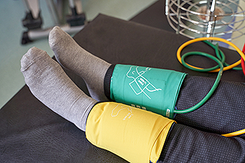
While PAD occurs in the legs and arteries, Podiatrists can diagnose PAD. Podiatrists utilize a test called an ankle-brachial index (ABI). An ABI test compares blood pressure in your arm to you ankle to see if any abnormality occurs. Ultrasound and imaging devices may also be used.
Treatment
Fortunately, lifestyle changes such as maintaining a healthy diet, exercising, managing cholesterol and blood sugar levels, and quitting smoking, can all treat PAD. Medications that prevent clots from occurring can be prescribed. Finally, in some cases, surgery may be recommended.
If you have any questions, please feel free to contact one of our offices located in Traverse City and Kalkaska, MI . We offer the newest diagnostic and treatment technologies for all your foot care needs.
Published in Blog
Tagged under
Tuesday, 12 March 2024 00:00
Peripheral Artery Disease
Peripheral artery disease (PAD), or peripheral arterial disease, is a circulatory problem in which there is a reduction of blood flow to the limbs due to narrowed arteries. When peripheral artery disease develops, the extremities do not receive enough blood flow; this may cause symptoms to develop such as claudication, or leg pain when walking. The legs are the most common site of peripheral artery disease.
Claudication, or leg pain when walking, is one of several symptoms that can develop due to peripheral artery disease. Other symptoms caused by the disease include painful cramping in the hips, thighs, or calves after certain activities; leg numbness or weakness; coldness in the lower leg or foot; sores on the lower extremities that do not heal; hair loss on the lower extremities; and a missing or weak pulse in the lower extremities. In more severe cases, pain may even occur when the body is at rest or when lying down.
Peripheral artery disease is typically caused by atherosclerosis, a condition in which fatty deposits build up in the arterial walls and reduce blood flow. Smoking, diabetes, obesity, high blood pressure, and high cholesterol are some of the risk factors for peripheral artery disease.
If you are experiencing pain, numbness, or other symptoms in the lower extremities, see your healthcare professional immediately. Diagnosed peripheral artery disease can be treated with various medications, angioplasty and surgery, exercise programs, or alternative medicine. It is important to consult a healthcare professional to determine the best treatment for you.
Published in Featured
Tagged under
Tuesday, 05 March 2024 00:00
Symptoms and Risk Factors of Bunions
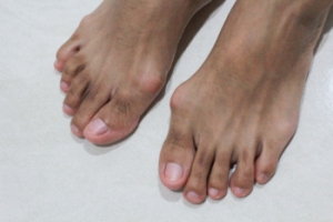
Bunions are common foot deformities that can be recognized by the formation of a bony bump at the base of the big toe. The tip of the toe is then pushed in the opposite direction, toward the smaller toes. A bunion is often accompanied by pain, inflammation, restricted toe movement, and the development of calluses or corns. While anyone can develop a bunion, certain risk factors heighten susceptibility. Wearing ill-fitting footwear, particularly high heels or narrow shoes, exerts pressure on the toes and contributes to bunion formation. Additionally, genetic predispositions play a significant role, with family history indicating a higher likelihood of developing foot structure abnormalities that may lead to bunions. Various foot conditions such as flat feet, low arches, or previous foot injuries increase the risk. Medical conditions like rheumatoid arthritis, gout, and connective tissue disorders are often associated with bunions. If you or your child show a tendency toward developing a bunion, it is suggested that you schedule an appointment with a podiatrist for an evaluation and recommended treatment measures.
If you are suffering from bunions, contact Dr. Ronald K. Olm of Grand Traverse Foot & Ankle Center. Our doctor can provide the care you need to keep you pain-free and on your feet.
What Is a Bunion?
A bunion is formed of swollen tissue or an enlargement of boney growth, usually located at the base joint of the toe that connects to the foot. The swelling occurs due to the bones in the big toe shifting inward, which impacts the other toes of the foot. This causes the area around the base of the big toe to become inflamed and painful.
Why Do Bunions Form?
Genetics – Susceptibility to bunions are often hereditary
Stress on the feet – Poorly fitted and uncomfortable footwear that places stress on feet, such as heels, can worsen existing bunions
How Are Bunions Diagnosed?
Doctors often perform two tests – blood tests and x-rays – when trying to diagnose bunions, especially in the early stages of development. Blood tests help determine if the foot pain is being caused by something else, such as arthritis, while x-rays provide a clear picture of your bone structure to your doctor.
How Are Bunions Treated?
- Refrain from wearing heels or similar shoes that cause discomfort
- Select wider shoes that can provide more comfort and reduce pain
- Anti-inflammatory and pain management drugs
- Orthotics or foot inserts
- Surgery
If you have any questions, please feel free to contact one of our offices located in Traverse City and Kalkaska, MI . We offer the newest diagnostic and treatment technologies for all your foot care needs.
Published in Blog
Tagged under
Tuesday, 05 March 2024 00:00
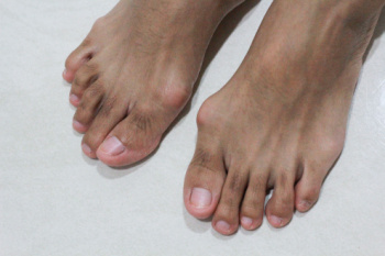
What Are Bunions?
Bunions are large bony bumps at the base of the big toe. Medically known as hallux valgus, a bunion is a misalignment of the metatarsophalangeal joint, or big toe joint. The misalignment will generally worsen with time if left untreated.
The exact cause of bunions is unknown, with genetics seen as a potential cause. High heels and poorly-fitted footwear, rheumatoid arthritis, and heredity all seem to be potential factors behind the exacerbation of bunions. Women have been found to be more likely to develop bunions in comparison to men.
Bunions do not always produce symptoms. The best way to tell is if the big toe is pushing up against the next toe and there is a large protrusion at the base of the big toe. You may or may not feel pain. Redness, swelling, and restricted movement of the big toe may be present as well.
Podiatrists use a variety of methods to diagnose bunions. If there are symptoms present, podiatrists will first consider that it is a bunion. If not, a physical examination will be conducted to check function of the big toe. Finally, an X-ray may be taken to view the extent of the bunion and confirm it is a bunion.
Typically, nonsurgical methods are used to treat bunions, unless the bunion has become too misaligned. Orthotics, icing and resting the foot, roomier and better fitted shoes, taping the foot, and pain medication are usually utilized first. If the bunion doesn’t go away or causes extreme pain, surgery may be required. Surgeons will either remove part of the swollen tissue or bone to straighten the toe out.
If you have a bunion, it is recommended to see a podiatrist. The longer it is left untreated, the worse it may get. Podiatrists can properly diagnose and treat a bunion before it gets worse.
Published in Featured
Tagged under
Friday, 01 March 2024 00:00
Gout Pain Can Be Managed


Gout is a painful, inflammatory form of arthritis. Those affected will typically feel an intense stiffness in the joints of their feet, particularly in the big toe. Schedule a visit to learn about how gout can be managed and treated.
Published in Blog
Tagged under
Tuesday, 27 February 2024 00:00
Managing Various Grades of Ankle Sprains
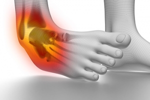
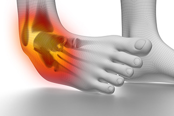
Ankle sprains are common injuries that can disrupt daily activities and sports participation. Effective management of ankle sprains involves understanding the severity of the injury and implementing appropriate treatment strategies. Ankle sprains are often classified into three grades, based on the extent of ligament damage. Grade I sprains involve mild stretching or microscopic tears of the ligaments, resulting in minimal swelling and pain. Grade II sprains involve partial tearing of the ligaments, leading to moderate swelling, pain, and instability. Grade III sprains are the most severe, involving complete tearing of the ligaments, significant swelling, and joint instability. Management of ankle sprains typically begins with elevation which may help to reduce any existing swelling. In severe cases, immobilization with a brace or boot may be necessary to facilitate healing. Gradual return to weight-bearing activities and sports should be guided by pain tolerance and functional improvement, aiming to prevent recurrent injuries and promote long-term ankle health. If you have sprained an ankle, it is suggested that you consult a podiatrist who can determine the grade of the sprain and offer appropriate treatment methods.
Although ankle sprains are common, they aren’t always minor injuries. If you need your ankle injury looked at, contact Dr. Ronald K. Olm from Grand Traverse Foot & Ankle Center. Our doctor can provide the care you need to keep you pain-free and on your feet.
How Does an Ankle Sprain Occur?
Ankle sprains are the result of a tear in the ligaments within the ankle. These injuries may happen when you make a rapid shifting movement while your foot is planted. A less common way to sprain your ankle is when your ankle rolls inward while your foot turns outward.
What Are the Symptoms?
- Pain at the sight of the tear
- Bruising/Swelling
- Ankle area is tender to touch
- In severe cases, may hear/feel something tear
- Skin discoloration
Preventing a Sprain
- Wearing appropriate shoes for the occasion
- Stretching before exercises and sports
- Knowing your limits
Treatment of a Sprain
In many cases, the RICE method (Rest, Ice, Compression, and Elevate) is used to treat ankle sprains. However, you should see a podiatrist to see which treatment option would work best with your injury. In severe cases, surgery may be required.
It is important to ask your doctor about rehab options after you receive treatment for your injury. Stretching, strength training, and balance exercises may help the ankle heal while also preventing further injury.
If you have any questions, please feel free to contact one of our offices located in Traverse City and Kalkaska, MI . We offer the newest diagnostic and treatment technologies for all your foot care needs.
Published in Blog
Tagged under
Tuesday, 27 February 2024 00:00
Three Grades of Ankle Sprains
An ankle sprain occurs when one or more ankle ligament gets overly stretched. Ligaments are strong bands of tissue that bind and support the bones and other structures that make up the ankle. In more severe ankle sprains, the ligament(s) tear—either partially or completely—and there may be an audible popping noise at the moment of injury.
Ankle sprains are quite common and can occur when the ankle rolls outwardly (eversion) or inwardly (inversion), causing the ligament(s) to stretch beyond normal limits, or even tear. Falls, twists, or blows to the ankle during sports or other activities can cause this injury, as well as wearing improper footwear, running on uneven surfaces, or having weak ankles.
Depending on the injury’s severity, an ankle sprain will be classified as Grade I, Grade II, or Grade III. Grade I sprains involve ligament(s) being overly stretched but not torn, with symptoms of mild pain, swelling, and ankle instability. There may also be some difficulty bearing weight. A Grade II sprain usually involves a partial tear of the ligament which brings more intensity in these symptoms, along with possible bruising. With a Grade III sprain, the ligament is completely torn, the symptoms are severe, and it may not be possible to put weight on the affected foot at all.
To diagnose and grade an ankle sprain, a podiatrist will perform a physical examination, checking for tenderness and range of motion in the ankle. For more severe sprains, X-rays or other imaging studies may be necessary.
It is vitally important to have an ankle sprain treated properly as improper healing often leads to future ankle sprains and possibly even chronic ankle stability. Treatment for an ankle sprain will vary, depending on its severity, and may include the RICE method (Rest/Ice/Compression/Elevation), physical therapy, bracing, medications, and possibly even surgery to repair a torn ligament. Rehabilitation is very important for the sprain to heal properly and to restore functionality.
Published in Featured
Tagged under
Tuesday, 20 February 2024 00:00
Past, Present, and Future of Diabetic Foot Ulcer Treatment

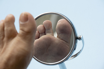
The surge in global diabetes has led to a rise in diabetic foot ulcers, posing significant health challenges. In the mid-19th century, Marchal de Calvi and Thomas Hodgkin identified the link between diabetes and foot gangrene, paving the way for pioneering treatments like Frederick Treves' sharp debridement method. Treves emphasized pressure relief and patient education. The discovery of insulin in the 20th century improved diabetic patients' lives but also increased foot disease cases. Elliot Joslin established the first foot clinic in 1928, advocating a multidisciplinary approach. Penicillin's discovery in 1928 revolutionized infection treatment, reducing amputation rates. Revascularization techniques and limb salvage became critical, with Frank Wheelock pioneering bypass grafts. Topical negative pressure therapy emerged in the 1990s, aiding wound healing. Despite advancements, chronic diabetic ulcers remain a challenge, with ongoing research for new treatments. Current treatment involves a multidisciplinary approach, controlling diabetes, and utilizing various wound care methods. Challenges persist, underscoring the importance of diabetics scheduling routine appointments with a podiatrist for foot examinations and specialized care.
Wound care is an important part in dealing with diabetes. If you have diabetes and a foot wound or would like more information about wound care for diabetics, consult with Dr. Ronald K. Olm from Grand Traverse Foot & Ankle Center. Our doctor will assess your condition and provide you with quality foot and ankle treatment.
What Is Wound Care?
Wound care is the practice of taking proper care of a wound. This can range from the smallest to the largest of wounds. While everyone can benefit from proper wound care, it is much more important for diabetics. Diabetics often suffer from poor blood circulation which causes wounds to heal much slower than they would in a non-diabetic.
What Is the Importance of Wound Care?
While it may not seem apparent with small ulcers on the foot, for diabetics, any size ulcer can become infected. Diabetics often also suffer from neuropathy, or nerve loss. This means they might not even feel when they have an ulcer on their foot. If the wound becomes severely infected, amputation may be necessary. Therefore, it is of the upmost importance to properly care for any and all foot wounds.
How to Care for Wounds
The best way to care for foot wounds is to prevent them. For diabetics, this means daily inspections of the feet for any signs of abnormalities or ulcers. It is also recommended to see a podiatrist several times a year for a foot inspection. If you do have an ulcer, run the wound under water to clear dirt from the wound; then apply antibiotic ointment to the wound and cover with a bandage. Bandages should be changed daily and keeping pressure off the wound is smart. It is advised to see a podiatrist, who can keep an eye on it.
If you have any questions, please feel free to contact one of our offices located in Traverse City and Kalkaska, MI . We offer the newest diagnostic and treatment technologies for all your foot care needs.
Published in Blog
Tagged under
Tuesday, 20 February 2024 00:00
Wound Care
Diabetics must be wary of all wounds, regardless of depth or size. Diabetes, a chronic disease in which the body cannot properly use glucose the way it normally would, causes various complications that make wounds difficult to heal. Nerve damage or neuropathy will cause diabetics to have trouble feeling the pain of a blister or cut until the condition has significantly worsened or become infected. A diabetic’s weakened immune system can make even the most minor of wounds easily susceptible to infection. Diabetics are also more prone to developing narrow, clogged arteries, and are therefore more likely to develop wounds.
Wounds should be taken care of immediately after discovery, as even the smallest of wounds can become infected if enough bacteria build up within the wound. To remove dirt, wounds should be first rinsed under running water only. Soap, hydrogen peroxide, or iodine can irritate the injury and should be avoided. To prevent infection, apply antibiotic ointment to the wound and cover it with a bandage. The bandage should be changed daily. The skin around the wound may be cleaned with soap.
To prevent further exacerbation, see a doctor—especially if you have diabetes. Minor skin conditions can become larger problems if not properly inspected. As the wound heals, make sure to avoid applying pressure to the affected area.
Published in Featured
Tagged under